Currently I work as the osteoarchaeologist for the COMMIOS project at the University of York. As part of this project I analyse human remains from the British Iron Age. I am hugely curious about every skeleton that I encounter, and I love teasing as much information out of every bone and fragment as possible. In this blog I will tell you about the work I do, how I analyse a skeleton and the information we can gain from the human remains.
The first step in identifying an Iron Age individual is to construct the ‘biological profile’. This is done by analysing the skeleton to provide information about age, sex and stature as well as additional information of trauma and disease. This provides the life course history of the individual (from birth to death), from the osteoarchaeological evidence of the human remains themselves.
How you approach a skeletal analysis depends on their context – is this an isolated bone? Are they comingled and fragmented remains from a mortuary cave? Is it a mass burial or a single burial (i.e. an ‘inhumation’) or is it a cremation? The first step is to assess whether there are only human remains present (rather than animal remains), and if there is one or more individuals and separate these out. So, let’s presume we have done that, and move forward with the skeletal analysis of a single inhumation. The next step is to determine if it is a child (i.e. a ‘non-adult’) or an adult, as the assessment of age uses different methods. If it is a child, we do not assess sex, as the sexual characteristics do not start to be expressed until puberty and are rarely clear enough until late adolescence. Assessment of sex in adults is predominantly done using the pelvis and skull but measurements of other areas of the skeleton can also be used. As a response to the pubertal hormones, the female pelvis changes in preparation to being able to pass a child through the pelvic canal, so the male pelvis retains the more native shape. The opposite is true for the skull – the female skull retains the more native (child-like) appearance, while the male skull changes to a more masculine appearance. One of the key research aims for the COMMIOS project is to analyse skeletons for ancient DNA which means was can also determine the genetic sex of an individual.
Assessment of age from a child skeleton is much easier than in adults, as skeletal development during childhood is a hugely dynamic and continual process. The most precise indicator of age is dental development (i.e. ‘dental age’) as this is more genetically directed rather than being influenced by the environment.

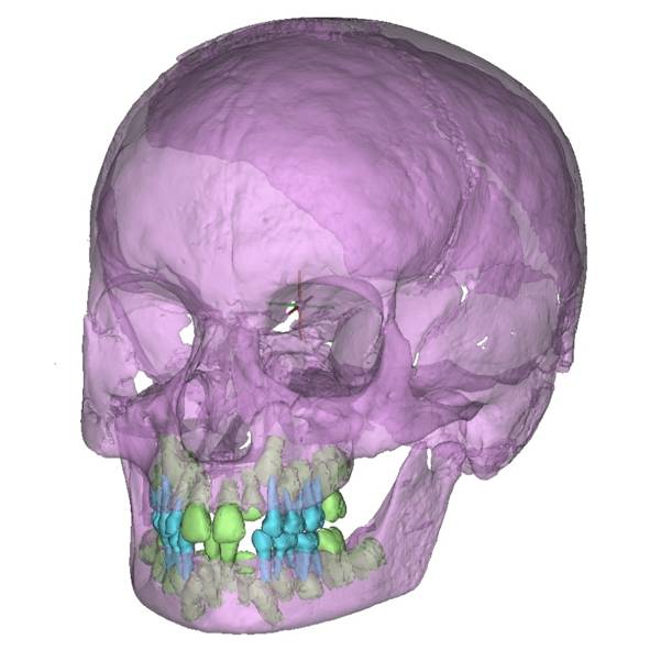
Figure 1: CT scan of an archaeological child’s skull showing dental development. Deciduous teeth (milk teeth) are coloured blue, permanent teeth are coloured green. Based on dental eruption, enamel formation and root development, age-at-death for this individual can be estimated to about seven years. Image: C. Primeau.
If there are no teeth available, the development of bones and fusion of skeletal elements can be used (i.e. ‘skeletal age’). Length of long bones can be used as a rough indicator of age as a last resort. However, the development of bones is more influenced by the environment, as development can be delayed due to periods of disease, malnutrition, or starvation. Also, length of long bones, which is essentially related to height, is also naturally highly variable between children. We have all seen a class of children of similar ages and how variable they are in their development; hence length of long bones is not very precise. The end of childhood and the commencement of adulthood is not clearly defined in osteoarchaeology, although the convention of less than/older than 18 years is often used. However, this exact age marker is difficult to assess skeletally, while osteologically the fusion of all long bones is a good marker of the end of skeletal growth. Younger adults can then be identified from a few late fusing skeletal elements such as the iliac crest (the top of the hip bones), the fusion between the two upper sacral vertebrae and the medial end of the clavicle. But once these skeletal elements have completely fused, there is far less information available to indicate age, apart from the very slow and highly variable degeneration of the bones and wear on teeth. Therefore, the age categories we use for adult skeletons are much wider than what we use for children.
Stature is the calculation of height, which is preferably done using the femur (thigh bone) or the tibia (shin bone) as these two bones contribute to height rather than bones from the upper limbs (arms), although these can be used. Although stature tends to be within a similar range for males and females it can also be used as a proxy for health between individuals and populations.
To complete the skeletal analysis, we also look at trauma, meaning the timing of an injury to a bone. The timing of trauma can be assessed as ante-mortem (before the time of death) from signs of healing, peri-mortem (around the time of death) or as post-mortem damage (after death). However, what is actually assessed is the quality of bone as ‘wet’, meaning still retaining organic material (collagen) or ‘dry’, meaning having lost the collagen content and only retaining the inorganic components. Peri-mortem trauma is when there is no healing evident on the bone from an ante-mortem trauma and neither is there evidence of dry fractures (indicating post-mortem fractures). However, this period can be several weeks on either side of death and not necessarily be related to circumstances surrounding the event of death, hence differentiating between peri-mortem and post-mortem trauma can be particularly challenging.
Lastly, we look at indicators of health on the skeleton. The teeth can provide evidence about childhood health as the enamel is formed during childhood and does not remodel after completion. Hence lines of disruption to the enamel formation provides glimpses into periods of a compromised health during the growing years. During later years, caries, gum disease and tooth loss provides further information of health. Later in life, an individual may develop degenerative changes visible on the skeleton, telling a tale of manual labour and physical hardship. Many infectious diseases will not leave evidence on a skeleton, and neither will many rapid causes of death such as septicaemia. Some infections will leave signs on the skeleton but often they are not specific enough for us to narrow down what type of infection was bothering the Iron Age person. A few infections can be more specifically identified on a skeleton, such as tuberculosis, although only a minority of people have tuberculosis that will be expressed skeletally. From such information we can piece together a life course history of an individual, from illnesses during earlier development and childhood, trauma and diseases during their adult life all the way through to their death. Collectively this information helps us build up a detailed understanding of the experiences of the individual.
And I’ll leave you with one final note – a skeletal analysis is only as good as the skeletal remains themselves! The more fragmented, degraded, and non-complete a skeleton is, the less information we can gather. The exciting part of working with skeletons from the Iron Age is the range of remains. The way people buried and treated their dead varied a lot in the Iron Age so I get to analyse a whole range of remains, such as isolated bones, non-complete remains, inhumations and cremations as well as a range of bone modifications, so as an osteologist the Iron Age keeps me on my toes!
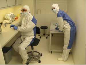
Figure 2: As the osteoarchaeologist for COMMIOS I also get involved in sampling material for aDNA, isotope and proteomics analysis. Here I am on the right, analysing a tooth in a clean lab, to avoid contamination prior to aDNA analysis. The image is from a project before COMMIOS.
This blog post was initially written for the COMMIOS project.