The Curious Case of the Forensic Strawberry
Exploring the playful side of micro-CT imaging
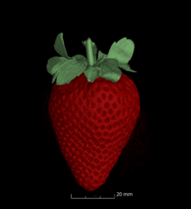
In forensic micro-CT, we’re usually concerned with some of the most serious aspects of human life—and death. We use high-resolution imaging to examine bones, tissues, and evidence in support of complex criminal investigations. But every once in a while, science calls for a change of pace. Enter: the strawberry.
This scan of a humble supermarket strawberry wasn’t part of any forensic casework. It wasn’t recovered from a crime scene or involved in any suspicious activity—unless you count going soft in the fridge. Instead, it was scanned purely for fun and as a way to showcase the astonishing level of anatomical detail that micro-CT can capture.
And it turns out, strawberries are fascinating.
Using the same forensic imaging setup we apply to post-mortem cases, the micro-CT scan revealed intricate internal structures: the distribution of seeds on the outer surface, internal air pockets, the vascular structure of the fruit, and even subtle density variations that tell us about its ripeness and texture. While these features aren’t exactly court-admissible evidence, they’re a great demonstration of what micro-CT can uncover—even in something as familiar as a piece of fruit.
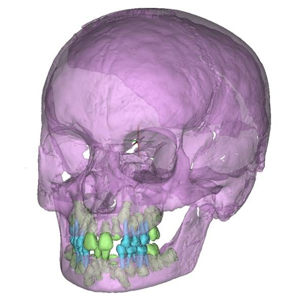
This light-hearted scan also has a serious purpose: science communication. Forensic imaging, particularly micro-CT, is often hidden from public view. It’s used in sensitive contexts such as homicides and abuse cases, where confidentiality and ethics rightly limit what we can show or discuss. But scanning a strawberry? That gives us an accessible, visually compelling way to talk about the power of this technology without compromising any real-world investigations.
It also sparks curiosity. Seeing an everyday object through a forensic lens can open the door to discussions about how micro-CT works, what it’s used for in practice, and why high-resolution imaging matters—not just in medicine or research, but also in criminal justice.