Welcome to my first guest blogger, my bone colleague: Annie Mason! I have invited Annie as her research combines two of my favorite subjects: radiographic images and animals, in Annie’s case primates – hope you enjoy!
Hi! I am Annie! I did my undergraduate degree in archaeology and anthropology at the University of Bristol. Towards the end of my degree, I spoke to Professor Kate Robson Brown about taking a Masters Degree. Upon hearing about my interests in primatology, bones and big data, Professor Robson Brown suggested a project that she had been considering and offered to supervise me. A previous student had digitised over one thousand film radiographs from Bristol Zoological Gardens, but had never performed analysis of the images. Professor Robson Brown suggested that they could be used for a study on bone development, and I was very excited to be given the opportunity to explore this! But why study bones of primates? As well as being interesting in their own right, they are our closest living relatives and therefore can inform our understanding of human evolution.
The first task was to organise the x-ray images from Bristol Zoo. Several were misfiled (including a fruit bat and a parrot, which were surprising to come across, see Figure 1a and b), or duplicated. Others did not have the necessary information, such as the date of the x-ray. This is important as the age of the primate on the x-ray can then be calculated, to the day.
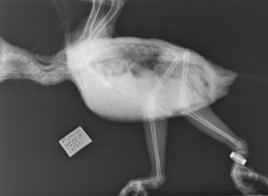
Figure 1a: Lateral view of a parrot. The endotracheal intubation tube is visible along the neck of the parrot. Reason for x-ray, sex and age are unknown.
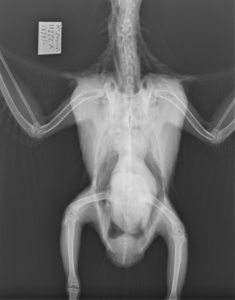
Figure 1b: Ventro-dorsal view of the same parrot as seen in Figure 1a, above. Part of the wing structure is faintly visible.
First, I selected the images from primates and then separated them into the 25 different primate species we had x-rays from. I was given access to a huge database used by zoos across the world, which allowed me to see the exact date of birth of each animal, and see whether they had suffered from any illness, which would have affected the growth of their skeletons. Then I started to look at bone development in relation to the age of each individual.
Radiographic images make longitudinal studies possible, when repeated x-rays of the same living primate are taken over short or long periods of time and then may be compared. This can be used for understanding how bones heal after a fracture (See Figure 2) in different species and at different ages of a primate’s life, and crucially for this study, how the bone changes as the individual matures.
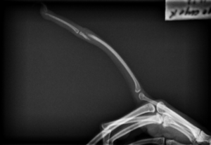
Figure 2: Lateral view of a hand of adult male Aye-Aye, nearly 5 years old, with a displaced fracture of the phalanx (finger bone). It is unknown how the Aye-Aye broke its finger.
So what growth changes can be seen in bones? Humans and other primates (a group including lemurs, lorises, tarsiers, monkeys, and apes) start life with a large number of their bones in three parts. Later these parts gradually fuse into a single bone. The fusion of the epiphyses (joint surfaces of the bones) to the diaphysis (main body, or shaft, of the bone) is called ‘epiphyseal fusion’. The end of this process marks the end of growth and the primate is now fully formed and has reached is full potential size. This process is used by archaeologists and anthropologists to work out how old an individual animal or person was at the time of death.
I then developed a method for ageing primates based on this bone development from radiographs, focusing on the long bones of the limbs (humerus, radius, femur, and tibia). These long bones were chosen as they were most often clearly seen in the radiographic images. My research developed a nine-stage scoring system in order to describe the formation and fusion of the epiphyses. I categorised the end of each bone as falling under stage 0 (where the epiphyses – end of the bone had not yet started to develop) to stage 8 (where there was no fusion line visible at all). See Figure 3a, 3b and 3c.
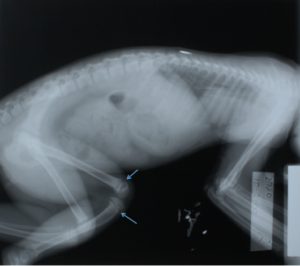
Figure 3a: Lateral view of a female Black Howler, age 9.5 months. Her epiphyses are not fused, which is most clearly seen in her knee joint (blue arrows).
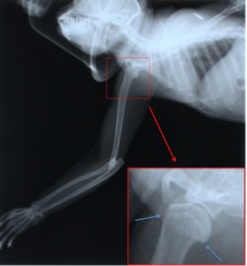
Figure 3b: Lateral view of a male Coppery Titi, age 2 years. The epiphyseal line is still visible on his upper arm bone (humerus) by his shoulder joint. The facemask to provide inhaled anesthetics is visible on the x-ray.

Figure 3c: Ventro-dorsal view of a male Gibbon, age nearly 16 years. His epiphyses on his thigh bone (femur) are clearly fused with no visible epiphyseal line by his hip joint (blue arrows).
Therefore, using my method, the age of the primate can be determined from a single x-ray much more accurately and further into adulthood, than with most previous research on the subject. It can be used to age an animal of unknown age as it enters a zoo collection (such as a rescue animal that cannot be rehabilitated) or to see if an animal has obtained age appropriate growth, or if it is experiencing developmental delay.
We see that in some species the continued radiographic visibility of the epiphyseal fusion line (See Figure 3b) can be used to determine the age of an individual well after the bones are fused. I also found that in the majority of species studied the proximal femur (hip joint, see Figure 3c) is the first long bone to reach full fusion. Why this is the case, I have not yet been able to answer. Perhaps the sequence of epiphyseal fusion may relate to locomotor styles (ways of moving around) of different species. Perhaps for some primate species, their locomotion means that the hip needs to be stronger at an earlier age.
Probably the most surprising finding of this study had to do with the adorable squirrel monkey. Both the males and females are the same size in adulthood, so you would expect them to grow at the same speed? Wrong! Some bones of the male skeleton, grow much faster than do in the female. (See Figure 4a and 4b). Again, this is something I have not yet been able to explain.
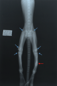
Figure 4a: Ventro-dorsal view of a female squirrel monkey, age 6.6 months old. Her epiphyses are clearly not fused, which is particularly visible in the knee and hip joints (blue arrows). On her lower left leg (red arrow), there is a bone lesion. The medical records did not clarify if this was an earlier fracture or a bone growth of unknown nature.
Figure 4b: Lateral view of a male squirrel monkey, age 6.4 months. The fusion of his knee and hip joints are more advanced than the female in Fig. 4a, despite the fact he is two months younger than her.
The hardest part of the study was that the radiographic collection included some images of an infant gorilla that were taken post-mortem (after death), see Figure 5. The similarities with a human baby were incredibly striking, which added to the sadness of seeing those x-rays.
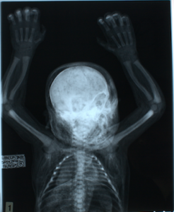
Figure 5: Post-mortem x-ray of an infant gorilla, ventro-dorsal view. Sex was not available to this study, age was two days old and cause of death was unknown.
On a more cheerful note, possibly the most fun fact about primate radiographs is, however, that the fantastic ears of the endangered Aye-Aye (another type of lemur) show up in their x-rays. See Figure 6a and 6b of an Aye-Aye, where the ears are clearly visible on the x-ray, showing their ear cartilage.
Figure 6a: Ventro-dorsal view of female age 14.5 years. The ear cartilage is clearly visible.
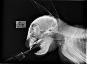
Figure 6b: Lateral view of same female from Fig. 6a. The ear cartilage is clearly visible.